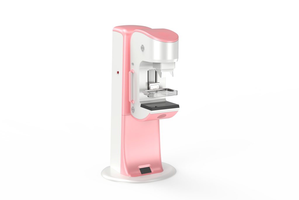
Breast cancer is one of the most common cancers affecting women worldwide and remains a leading cause of cancer related mortality. Early detection may plays a crucial role in improving survival rates and treatment outcomes. Among the available screening and diagnostic tools, mammography is considered the gold standard for the early detection of breast cancer.
This imaging technique has been widely used for decades and has proven effective in identifying breast abnormalities before clinical symptoms appear. Mammography is a specialized medical imaging technique that uses low dose X-rays to visualize the internal structure of the breast. It is designed to detect subtle changes in breast tissue, such as masses, calcifications, or architectural distortions, which may indicate the presence of cancer.
Mammography can be performed as:
- Screening mammography for asymptomatic women to detect cancer early
- Diagnostic mammography for evaluating specific breast complaints or abnormal screening results
Mammography plays a vital role in detecting breast cancer at an early stage, often before a lump can be felt during physical examination. Early-stage breast cancers are typically smaller, localized, and more treatable. Studies have shown that regular mammographic screening significantly reduces breast cancer mortality by enabling earlier diagnosis and timely intervention.
There are some key features detected by mammography include:
- Microcalcifications, which may be an early sign of ductal carcinoma in situ (DCIS)
- Breast masses with irregular borders
- Asymmetry or distortion of breast tissue
Mammography may offers several important advantages:
- Early detection: Identifies cancer before symptoms develop
- Reduced mortality: Associated with decreased breast cancer death rates
- Cost-effective: More affordable compared to advanced imaging techniques
- Wide availability: Accessible in many healthcare settings
Limitations of Mammography:
Despite its benefits, mammography has some limitations. Its sensitivity may be reduced in women with dense breast tissue, leading to false-negative results. Additionally, false-positive findings can occur, causing anxiety and unnecessary follow-up procedures. Exposure to low-dose radiation is another concern, although the risk is minimal compared to the benefits of early cancer detection.
Complementary Imaging Techniques:
To overcome some limitations, mammography is often combined with other imaging modalities such as breast ultrasound or magnetic resonance imaging (MRI), particularly in high-risk patients or those with dense breasts. These complementary tools can improve diagnostic accuracy and help guide biopsy decisions.
In conclusion, Mammography remains a cornerstone in the early detection of breast cancer and continues to play a critical role in reducing mortality associated with the disease. While it is not without limitations, its proven effectiveness, accessibility, and ability to detect cancer at an early stage make it an essential component of breast cancer screening programs. Ongoing advancements in imaging technology, such as digital and 3D mammography, are expected to further enhance its diagnostic performance and clinical value. (IW 1901)